国际肿瘤学杂志 ›› 2025, Vol. 52 ›› Issue (3): 144-151.doi: 10.3760/cma.j.cn371439-20241113-00022

• 论著 • 上一篇    下一篇

基于18F-FDG PET/CT原发灶影像组学的联合模型预测NSCLC淋巴结转移的价值

来瑞鹤1, 滕月1, 戎剑2, 盛丹丹3, 耿羽智3, 陈建新2, 蒋冲4, 丁重阳5, 周正扬6()   

  1. 1南京医科大学鼓楼临床医学院 南京鼓楼医院核医学科,南京 210008
    2南京邮电大学通信与信息工程学院宽带无线通信与传感网技术重点实验室,南京 210003
    3南京医科大学第二附属医院核医学科,南京 210011
    4四川大学华西医院核医学科,成都 610041
    5南京医科大学附属第一医院 江苏省人民医院核医学科,南京 210029
    6南京医科大学鼓楼临床医学院 南京鼓楼医院放射科,南京 210008
  • 收稿日期:2024-11-13 修回日期:2025-01-03 出版日期:2025-03-08 发布日期:2025-04-02
  • 通讯作者: 周正扬,Email:zyzhou@nju.edu.cn
  • 基金资助:
    南京市卫生科技发展专项资金(YKK24090)

Predictive value of a combined model for lymph node metastasis in NSCLC based on primary lesion radiomics from 18F-FDG PET/CT

Lai Ruihe1, Teng Yue1, Rong Jian2, Sheng Dandan3, Geng Yuzhi3, Chen Jianxin2, Jiang Chong4, Ding Chongyang5, Zhou Zhengyang6()   

  1. 1Department of Nuclear Medicine,Nanjing Drum Tower Hospital,Clinical Medical School of Nanjing Medical University,Nanjing 210008,China
    2Key Laboratory of Broadband Wireless Communication and Sensor Network Technology(Ministry of Education),School of Communications and Information Engineering,Nanjing University of Posts and Telecommunications,Nanjing 210003,China
    3Department of Nuclear Medicine,Second Affiliated Hospital of Nanjing Medical University,Nanjing 210011,China
    4Department of Nuclear Medicine,West China Hospital of Sichuan University,Chengdu 610041,China
    5Department of Nuclear Medicine,First Affiliated Hospital with Nanjing Medical University,Jiangsu Province Hospital,Nanjing 210029,China
    6Department of Radiology,Nanjing Drum Tower Hospital,Clinical Medical School of Nanjing Medical University,Nanjing 210008,China
  • Received:2024-11-13 Revised:2025-01-03 Online:2025-03-08 Published:2025-04-02
  • Contact: Zhou Zhengyang,Email:zyzhou@nju.edu.cn
  • Supported by:
    Nanjing Health Science and Technology Development Special Fund(YKK24090)

摘要:

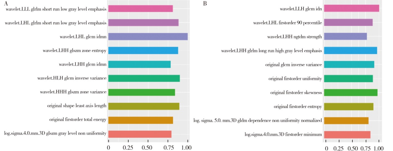
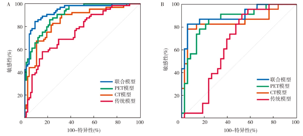
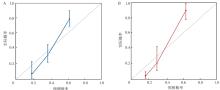
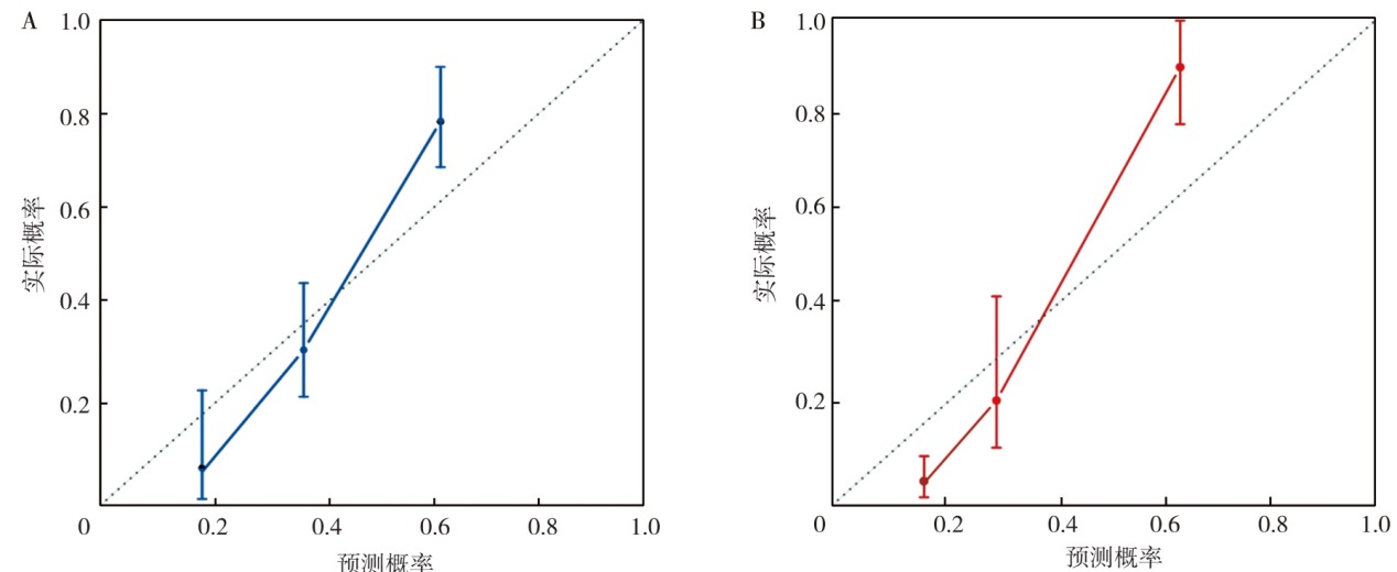
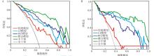
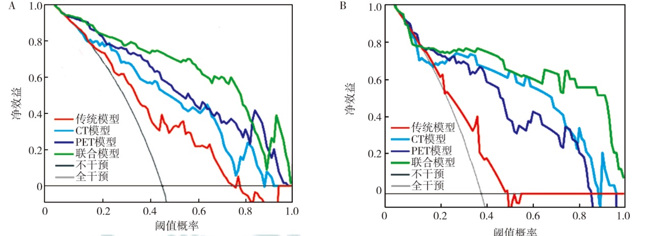
目的 评估基于原发灶18F-氟代脱氧葡萄糖(18F-FDG)PET/CT影像组学联合模型预测非小细胞肺癌(NSCLC)淋巴结转移的价值。方法 回顾性分析南京鼓楼医院2013年6月至2023年7月治疗前行PET/CT显像的203例NSCLC患者的临床资料。按照7∶3比例将患者随机分为训练集(n=142)和验证集(n=61)。在训练集中建立预测模型, 在训练集和验证集中分别对模型进行预测效能评估和临床应用价值验证。通过3D-slicer软件获得原发灶传统PET/CT参数和PET/CT影像组学特征。采用最小绝对收缩与选择算子(LASSO)、随机森林和极端梯度提升进行特征提取。采用支持向量机构建影像组学标签影像组学评分(Radscore)。采用单因素、多因素logistic回归分析预测NSCLC患者淋巴结转移的影响因素并建立模型, 采用受试者操作特征(ROC)曲线评估模型的预测效能, 采用校准曲线和临床决策曲线(DCA)评估模型的临床应用价值。结果 203例NSCLC患者中淋巴结转移116例, 其中训练集64例、验证集52例。采用3种互补的经典机器学习方法进行特征筛选, 最终分别得到10个影像组学特征。Radscore-PET的最佳阈值为0.43, Radscore-CT的最佳阈值为0.39。单因素分析显示, 性别(OR=0.48, 95%CI为0.24~0.95, P=0.036)、肿瘤标志物水平(OR=3.81, 95%CI为1.84~7.91, P<0.001)、肿瘤长径(OR=2.56, 95%CI为1.27~5.16, P=0.009)、肿瘤短径(OR=3.73, 95%CI为1.75~7.92, P=0.001)、空泡征(OR=0.32, 95%CI为0.12~0.86, P=0.024)、环形代谢(OR=3.67, 95%CI为1.33~10.13, P=0.012)、最大标准化摄取值(SUVmax)(OR=6.57, 95%CI为3.03~14.25, P<0.001)、肿瘤代谢体积(MTV)(OR=2.91, 95%CI为1.43~5.92, P=0.003)、病灶糖酵解总量(TLG)(OR=4.23, 95%CI为2.08~8.59, P<0.001)、Radscore-PET(OR=21.93, 95%CI为9.04~53.20, P<0.001)和Radscore-CT(OR=13.72, 95%CI为6.12~30.76, P<0.001)均是预测NSCLC患者淋巴结转移的影响因素。多因素分析显示, 肿瘤标志物水平(OR=2.55, 95%CI为1.11~5.90, P=0.028)、空泡征(OR=0.26, 95%CI为0.08~0.83, P=0.023)、SUVmaxOR=5.94, 95%CI为1.99~17.75, P=0.001)、Radscore-PET(OR=25.51, 95%CI为5.92~110.22, P<0.001)、Radscore-CT(OR=8.68, 95%CI为2.73~27.61, P<0.001)均是预测NSCLC患者淋巴结转移的独立影响因素。基于上述独立影响因素建立模型:传统模型(肿瘤标志物水平、空泡征、SUVmax)、PET模型(SUVmax、Radscore-PET)、CT模型(空泡征、Radscore-CT)和联合模型(肿瘤标志物水平、空泡征、SUVmax、Radscore-PET、Radscore-CT)。ROC曲线分析显示, 训练集中传统模型、PET模型、CT模型和联合模型的曲线下面积(AUC)分别为0.75(95%CI为0.67~0.82)、0.90(95%CI为0.84~0.95)、0.85(95%CI为0.78~0.90)和0.94(95%CI为0.88~0.97), 联合模型的预测价值高于传统模型(Z=5.01, P<0.001)、PET模型(Z=1.99, P=0.047)、CT模型(Z=3.25, P=0.001);验证集中传统模型、PET模型、CT模型和联合模型的AUC分别为0.65(95%CI为0.52~0.77)、0.86(95%CI为0.74~0.93)、0.85(95%CI为0.73~0.93)和0.90(95%CI为0.80~0.96), 联合模型的预测价值高于传统模型(Z=3.23, P=0.001)。训练集联合模型的敏感性和特异性分别为84.37%和91.03%, 验证集联合模型的敏感性和特异性分别为82.61%和94.74%。校准曲线显示, 训练集和验证集实际发生概率均与预测概率较为一致。DCA显示, 联合模型的辨别能力在训练集和验证集中均较好。结论 肿瘤标志物水平、空泡征、SUVmax、Radscore-PET、Radscore-CT均是预测NSCLC患者淋巴结转移的独立影响因素, 基于上述指标构建的联合模型对NSCLC淋巴结转移具有良好的预测效能和临床应用价值。

关键词: 癌, 非小细胞肺, 正电子发射断层显像计算机体层摄影术, 影像组学, 淋巴结转移

Abstract:

Objective To evaluate the value of a combined model based on primary lesion 18F-fluorodeoxyglucose(18F-FDG) PET/CT radiomics for predicting lymph node metastasis in non-small cell lung cancer(NSCLC). Methods A retrospective analysis was conducted on the clinical data of 203 NSCLC patients who underwent pre-treatment PET/CT imaging at Nanjing Drum Tower Hospital from June 2013 to July 2023. Patients were randomly assigned to the training set(n=142) and the validation set(n=61) at a ratio of 7∶3. A predictive model was developed in the training set, and its predictive performance and clinical application value were assessed in both the training and validation sets. Traditional PET/CT parameters and PET/CT radiomics features of the primary lesion were obtained by 3D-slicer software. Least absolute shrinkage and selection operator(LASSO), random forest, and extreme gradient boosting were performed to extract features. Support vector machine was used to construct a radiomics score(Radscore). Univariate and multivariate logistic regression analysis was used to predict the influencing factors of lymph node metastasis in NSCLC patients and to establish models. Predictive performance of the models was evaluated by receiver operator characteristic(ROC) curves and clinical application value was assessed by calibration curves and decision curve analysis(DCA). Results Among 203 NSCLC patients, 116 had lymph node metastasis, with 64 cases in the training set and 52 cases in the validation set. Three complementary classical machine learning methods were used for feature screening, and finally 10 radiomics features were obtained. The optimal threshold for Radscore-PET was 0.43 and the optimal threshold for Radscore-CT was 0.39. Univariate analysis showed that, sex(OR=0.48, 95%CI:0.24-0.95, P=0.036), tumor marker levels(OR=3.81, 95%CI:1.84-7.91, P<0.001), long diameter of tumor(OR=2.56, 95%CI:1.27-5.16, P=0.009), short diameter of tumor(OR=3.73, 95%CI:1.75-7.92, P=0.001), vacuolar sign(OR=0.32, 95%CI:0.12-0.86, P=0.024), ring-like metabolism(OR=3.67, 95%CI:1.33-10.13, P=0.012), maximum standardized uptake value(SUVmax)(OR=6.57, 95%CI:3.03-14.25, P<0.001), metabolic tumor volume(MTV)(OR=2.91, 95%CI:1.43-5.92, P=0.003), total lesion glycolysis(TLG)(OR=4.23, 95%CI:2.08-8.59, P<0.001), Radscore-PET(OR=21.93, 95%CI:9.04-53.20, P<0.001) and Radscore-CT(OR=13.72, 95%CI:6.12-30.76, P<0.001) were all influencing factors for predicting lymph node metastasis in NSCLC patients. Multivariate analysis showed that, tumor marker levels(OR=2.55, 95%CI:1.11-5.90, P=0.028), vacuolar sign(OR=0.26, 95%CI:0.08-0.83, P=0.023), SUVmaxOR=5.94, 95%CI:1.99-17.75, P=0.001), Radscore-PET(OR=25.51, 95%CI:5.92-110.22, P<0.001), and Radscore-CT(OR=8.68, 95%CI:2.73-27.61, P<0.001) were independent influencing factors for predicting lymph node metastasis in patients with NSCLC. Based on the above independent influencing factors, models were constructed:the traditional model(tumor marker levels, vacuolar sign, SUVmax), the PET model(SUVmax, Radscore-PET), the CT model(vacuolar sign, Radscore-CT), and the combined model(tumor marker levels, vacuolar sign, SUVmax, Radscore-PET, Radscore-CT). ROC curve analysis showed that, the area under curve(AUC) of the traditional, PET, CT, and combined models in the training set were 0.75(95%CI:0.67-0.82), 0.90(95%CI:0.84-0.95), 0.85(95%CI:0.78-0.90), and 0.94(95%CI:0.88-0.97), respectively. The predictive value of the combined model was higher than that of the traditional model(Z=5.01, P<0.001), the PET model(Z=1.99, P=0.047), and the CT model(Z=3.25, P=0.001). In the validation set, the AUCs for the traditional model, PET model, CT model, and combined model were 0.65(95%CI:0.52-0.77), 0.86(95%CI:0.74-0.93), 0.85(95%CI:0.73-0.93), and 0.90(95%CI:0.80-0.96), respectively. The predictive value of the combined model was superior to that of the traditional model(Z=3.23, P=0.001). The sensitivity and specificity of the combined model in the training set were 84.37% and 91.03%, while in the validation set, the sensitivity and specificity were 82.61% and 94.74%, respectively. Calibration curves showed a good agreement between the predicted and actual probabilities in both the training and validation sets. DCA showed that the combined models had good discriminative ability in both the training and validation sets. Conclusions Tumor marker levels, vacuolar sign, SUVmax, Radscore-PET, and Radscore-CT are all independent influencing factors for predicting lymph node metastasis in patients with NSCLC. The combined model based on these factors demonstrates excellent predictive performance and clinical application value for predicting lymph node metastasis in NSCLC.

Key words: Carcinoma, non-small-cell lung, Positron emission tomography computed tomography, Radiomics, Lymph node metastasis

Baidu
map