国际肿瘤学杂志 ›› 2025, Vol. 52 ›› Issue (3): 136-143.doi: 10.3760/cma.j.cn371439-20241021-00021

• 论著 • 上一篇    下一篇

外周实性结节Ⅰ期肺腺癌脏层胸膜侵犯的CT特征分析及预测价值

韩双()   

  1. 贵州中医药大学第二附属医院放射科,贵阳 550003
  • 收稿日期:2024-10-21 修回日期:2024-12-19 出版日期:2025-03-08 发布日期:2025-04-02
  • 通讯作者: Email:1527698862@qq.com

CT feature analysis and predictive value of visceral pleural invasion in stage Ⅰ lung adenocarcinoma with peripheral solid nodules

Han Shuang()   

  1. Department of Radiology,Second Affiliated Hospital of Guizhou University of Traditional Chinese Medicine,Guiyang 550003,China
  • Received:2024-10-21 Revised:2024-12-19 Online:2025-03-08 Published:2025-04-02
  • Contact: Email:1527698862@qq.com

摘要:

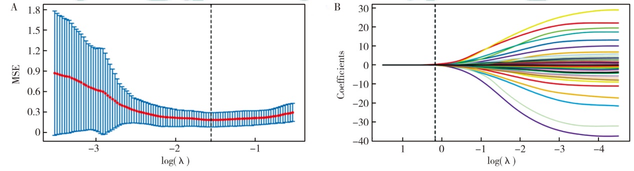
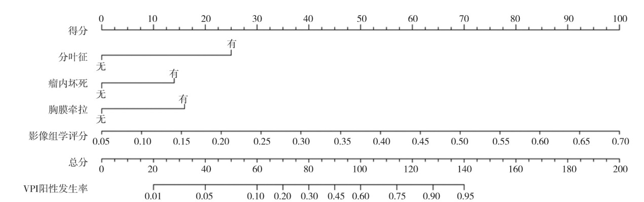
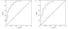
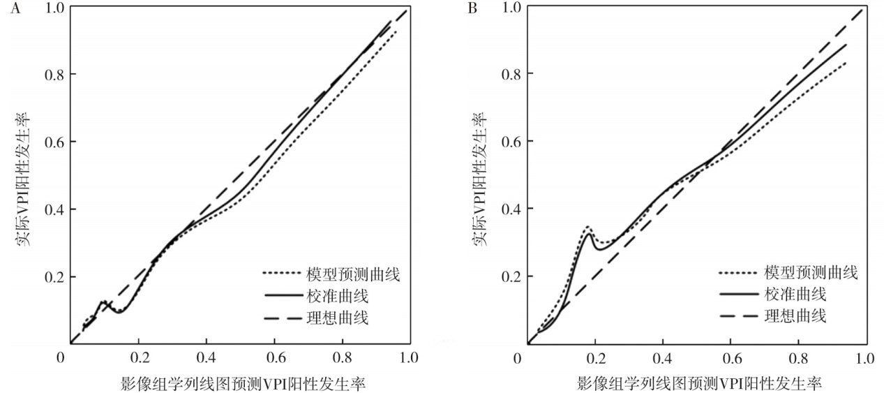
目的 探究外周实性结节Ⅰ期肺腺癌脏层胸膜侵犯(VPI)的CT特征及影像组学列线图的预测价值。方法 选择2022年8月至2023年11月贵州中医药大学第二附属医院收治的150例外周实性结节Ⅰ期肺腺癌患者为研究对象, 将2022年8月至2023年3月的患者定义为训练集(n=112), 2023年4月至11月的患者定义为验证集(n=38)。训练集用于建立模型, 训练集、验证集中分别进行模型性能评估。训练集中, 根据VPI发生情况, 将患者分为VPI阳性组(n=35)和VPI阴性组(n=77)。采用最小绝对收缩与选择算子(LASSO)回归分析对特征进行降维。采用多因素logistic回归分析预测VPI的影响因素, 根据多因素分析结果构建影像组学列线图预测模型。采用受试者操作特征(ROC)曲线及校准曲线评估预测模型的预测效能。结果 训练集VPI阴性组与阳性组患者的病理类型(χ2=11.49, P=0.003)、病灶最大径(t=5.83, P<0.001)、分叶征(χ2=9.29, P=0.002)、密度(χ2=8.32, P=0.004)、瘤内坏死(χ2=5.86, P=0.015)、胸膜牵拉(χ2=12.88, P<0.001)、胸膜接触(χ2=4.82, P=0.028)、邻近胸膜增厚(χ2=4.87, P=0.027)差异均有统计学意义。LASSO回归分析最终筛选出8个特征, 根据特征对应系数构建影像组学评分。单因素分析显示, 病灶最大径(OR=1.48, 95%CI为1.09~2.01, P=0.010)、分叶征(OR=5.09, 95%CI为2.31~6.00, P=0.001)、密度(OR=4.25, 95%CI为1.47~7.18, P=0.004)、瘤内坏死(OR=2.27, 95%CI为1.01~5.17, P=0.049)、胸膜牵拉(OR=6.75, 95%CI为1.92~13.68, P<0.001)、胸膜接触(OR=3.58, 95%CI为1.18~5.65, P=0.018)、邻近胸膜增厚(OR=3.60, 95%CI为1.18~5.72, P=0.018)、影像组学评分(OR=19 418.06, 95%CI为394.18~957 161.04, P<0.001)均是预测外周实性结节Ⅰ期肺腺癌患者VPI的影响因素。多因素分析显示, 分叶征(OR=6.42, 95%CI为1.42~18.58, P=0.018)、瘤内坏死(OR=3.63, 95%CI为1.01~10.01, P=0.046)、胸膜牵拉(OR=4.19, 95%CI为1.17~10.92, P=0.028)、影像组学评分(OR=179 711.20, 95%CI为525.13~61 552 573.59, P<0.001)均是预测外周实性结节Ⅰ期肺腺癌患者VPI的独立影响因素。将多因素分析中有统计学意义的指标建立影像组学列线图预测模型。ROC曲线分析显示, 训练集和验证集中, 影像组学列线图模型预测外周实性结节Ⅰ期肺腺癌患者VPI的曲线下面积(AUC)分别为0.88(95%CI为0.82~0.94)和0.87(95%CI为0.78~0.97), 敏感性分别为93%、82%, 特异性分别为72%、80%。训练集和验证集的C-index分别为0.89(95%CI为0.84~0.96)和0.88(95%CI为0.78~0.99), 两集的校准曲线均与理想曲线拟合良好。结论 外周实性结节Ⅰ期肺腺癌VPI的CT特征为有分叶征、瘤内坏死、胸膜牵拉, 基于CT特征的分叶征、瘤内坏死、胸膜牵拉、影像组学评分构建的影像组学列线图预测模型对外周实性结节Ⅰ期肺腺癌患者VPI具有较高的预测效能。

关键词: 肺腺癌, 胸膜, 列线图, 影像组学

Abstract:

Objective To explore the CT features and predictive value of radiomics nomogram of visceral pleural invasion (VPI) in stage Ⅰ lung adenocarcinoma with peripheral solid nodules. Methods One hundred and fifty patients with stage Ⅰ lung adenocarcinoma with peripheral solid nodules treated at the Second Affiliated Hospital of Guizhou University of Traditional Chinese Medicine from August 2022 to November 2023 were selected as the study objects. Patients from August 2022 to March 2023 were defined as the training set (n=112), and patients from April 2023 to November 2023 were defined as the validation set (n=38). The training set was used to build the model, and the training set and validation set were used to evaluate the model performance respectively. In the training set, patients were divided into VPI positive group (n=35) and VPI negative group (n=77) based on the occurrence of VPI. Least absolute shrinkage and selection operator (LASSO) regression analysis was used to reduce the dimensionality of features. Multivariate logistic regression analysis was used to predict the influencing factors of VPI, and a radiomics nomogram prediction model was constructed based on the results of the multivariate analysis. Receiver operator characteristic (ROC) curves and calibration curves were used to evaluate the predictive efficacy of the prediction model. Results There were statistically significant differences in pathological types (χ2=11.49, P=0.003), focal maximum diameter (t=5.83, P<0.001), lobulation sign (χ2=9.29, P=0.002), density (χ2=8.32, P=0.004), intratumoral necrosis (χ2=5.86, P=0.015), pleural traction (χ2=12.88, P<0.001), pleural contact (χ2=4.82, P=0.028), and adjacent pleural thickening (χ2=4.87, P=0.027) between the VPI positive group and negative group in the training set. LASSO regression analysis showed that 8 features were ultimately selected, and radiomics scores were constructed based on the corresponding coefficients of the features. Univariate analysis showed that, focal maximum diameter (OR=1.48, 95%CI:1.09-2.01, P=0.010), lobulation sign (OR=5.09, 95%CI:2.31-6.00, P=0.001), density (OR=4.25, 95%CI:1.47-7.18, P=0.004), intratumoral necrosis (OR=2.27, 95%CI:1.01-5.17, P=0.049), pleural traction (OR=6.75, 95%CI:1.92-13.68, P<0.001), pleural contact (OR=3.58, 95%CI:1.18-5.65, P=0.018), adjacent pleural thickening (OR=3.60, 95%CI:1.18-5.72, P=0.018), and radiomics score (OR=19 418.06, 95%CI:394.18-957 161.04, P<0.001) were all influencing factors in the prediction of VPI in peripheral solid nodule stage Ⅰ lung adenocarcinoma patients. Multivariate analysis showed that, lobulation sign (OR=6.42, 95%CI:1.42-18.58, P=0.018), intratumoral necrosis (OR=3.63, 95%CI:1.01-10.01, P=0.046), pleural traction (OR=4.19, 95%CI:1.17-10.92, P=0.028), and radiomics score (OR=179 711.20, 95%CI:525.13-61 552 573.59, P<0.001) were independent influencing factors in the prediction of VPI in patients with stage Ⅰ peripheral solid nodules of lung adenocarcinoma. A radiomics nomogram prediction model was established for indicators with statistical significance in multivariate analysis. ROC curve analysis showed that in the training set and validation set, the area under the curve (AUC) of the radiomics nomogram model predicting VPI of patients with peripheral solid nodules in stage Ⅰ lung adenocarcinoma was 0.88 (95%CI:0.82-0.94) and 0.87 (95%CI:0.78-0.97), respectively, and the sensitivity was 93% and 82%, respectively. The specificity was 72% and 80%, respectively. C-indices of the training and validation set were 0.89 (95%CI:0.84-0.96) and 0.88 (95%CI:0.78-0.99), respectively, and the calibration curves of both sets fitted well with the ideal curve. Conclusions The CT features of VPI in stage Ⅰ lung adenocarcinoma with peripheral solid nodules are lobular sign, intratumoral necrosis, and pleural traction. The radiomics nomogram model based on CT features of lobular sign, intratumoral necrosis, pleural traction, and radiomics score can predict VPI in patients with peripheral solid nodules in stage Ⅰ lung adenocarcinoma has high predictive efficacy.

Key words: Adenocarcinoma of lung, Pleura, Nomograms, Radiomics

Baidu
map